Gavin Brink
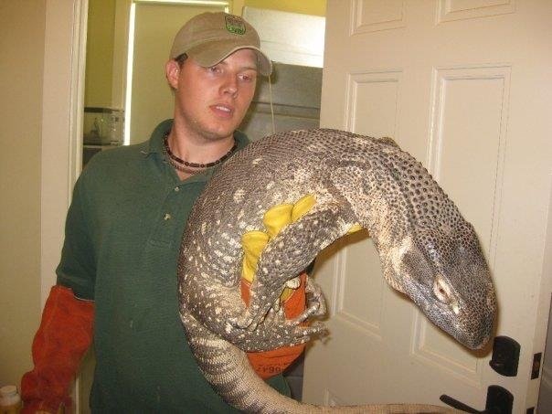
Gavin was a devoted herpetologist whose life was taken too soon. His family is continuing his legacy through the Gavin Brink Memorial Fund and through a property in Costa Rica, The Camaquiri Conservation Initiative (www.camaquiri.com).
Gavin got his undergraduate degree at Illinois State University and continued his education for a degree in Biology and Education at Northern Illinois University. He founded numerous businesses helping animals - a herpetology education station at a Wildlife Museum and a mobile veterinary business. He was devoted to teaching kids in a friendly and calm demeanor, putting everyone at ease around some creatures they may usually fear.
A beautiful tribute made by Gavin’s friend, Jessica Wadleigh.

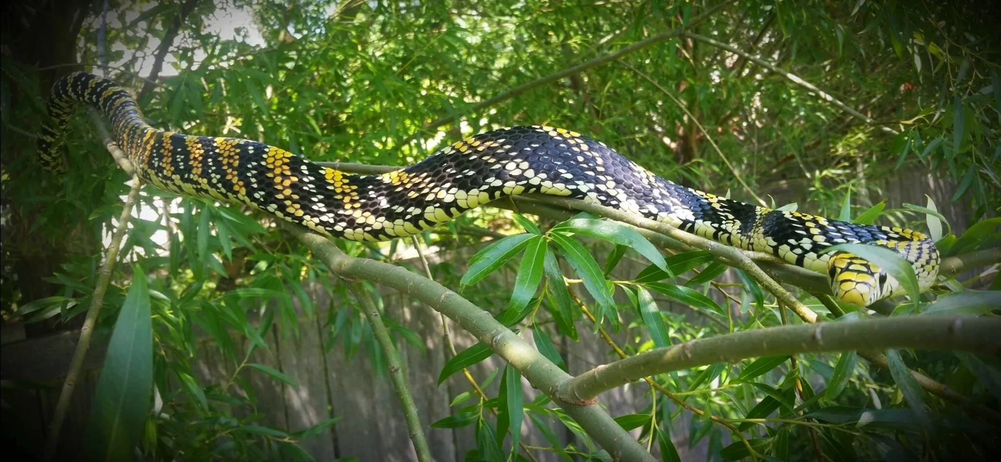
Near the end of 2018, the reptile community came together to celebrate the life of a dear friend, who left us too soon. Gathered at the celebration, nearly 300 people signed the guestbook in a very cramped bar and restaurant in Sycamore, Illinois. There were certainly more there who didn’t sign, and countless others who wished they could attend. Pictures were found on every table, free tequila shots on another, videos playing overhead of his antics, and a beautiful cake decorated to look like his favorite species, Spilotes pullatus mexi- canus, or the Mexican tiger rat snake. I stumbled across one of our mutual friends, an instructor at NIU from our Tropical Herpetology class in Costa Rica, who humbly said to me, “I don’t think I’ve spoken to this many people in my entire life.” It was a party, exactly as he would have wanted. Because you see, Gavin Brink hated funerals. He spoke about it adamantly whenever it came up. He hated the sad atmosphere, where he believed someone’s life should be celebrated. His family lovingly provided him with that celebration.
But he did love parties. Gavin was incredibly well known within the reptile community, partially because of his ability to bring people together. He would host annual “That’s Texas” parties, which involved tongue-in-cheek Texas-themed events, not limited to boxing over a Texas flag painted on the lawn, slip-n-slides, tequila fountains, and even a rented mechanical bull. He would often be the center of attention at the semi-annual NARBC Tinley shows, sometimes having a room in the hotel where people would gather as he made his classic “Prison Sen- tence” beverage, a horrible concoction of tequila, 90% grain alcohol infused with the hottest peppers he could find, MSG coated rims, and homeopathic pills made with trace amounts of Bothrops venom. But somehow, it was always the happening place. He would tell every table where to go at the show, and people from all walks of life would come. The more people the happier he was.
Perhaps it wasn’t the parties that made him happy, but bringing people together. Despite his often eccentric gestures, he always succeeded in winning people’s hearts as that strange but incredibly lovable brother figure. There are few others in the community with as far of a reach as he had. His connections extended anywhere that had anything to do with reptiles. With his zoological exploits, academia, the CHS, Costa Rica, his trolling-but- educational anonymous online presence as “Mace Montana” in countless Facebook groups, and the commu- nity of breeders of unique and otherwise unrecognized species of colubrids---no matter where you went in the reptile community, if you mentioned Gavin Brink or Mace Montana, someone would recognize the name.
He was a man of knowledge and learning as well. He would always try to educate, without ever patronizing anyone for their ignorance or misconceptions---particu- larly when it came to the topic of poison- ous snakes (yes, poisonous), on which he did a talk for the CHS in 2015. He loved working with kids, even doing a Junior Herpers talk and teaching children how to safely hook non-venomous snakes. Animals were certainly his passion, but he also wanted to spread that passion with everyone that he could.
Costa Rica, on the other hand, was where his spirit truly came alive. He taught as an assistant instructor in the Tropical Herpetology course I attended two years in a row. A class that was inadvisable for him because of his health, but that he refused to miss out on anyway. He’d been there many years prior to when I had the chance to go with him, but those memories will al- ways be dear to me. In the dark of primary rainforest, we belted out ridiculous songs, just to keep spirits up as our exhaustion took over from the difficult 9-mile hike through mud as deep as our thighs. At one point, one of my boots was caught in a cow track, the suction pulling the ill-fitted boot off my foot. Another student and I, who often brought up the rear of the hikes, found ourselves trapped in the mud attempting to free it. He returned, laughing and taking pictures of our muddy situation before helping us back out, insisting that we cannot allow the mud gods to take such a sacrifice. We found many herps that year and the year to follow, toasting the end to our long nights with Imperial and tequila. Truly, though, I feel that I was able to glimpse his true self, in a way that you can only see in the depths of the Costa Rican rain- forest.
At the most recent That’s Texas, he spoke to me at length. My partner and I hadn’t been able to come in a few years because of the timing of obligations, so he expressed how happy he was that we came and in his words, “I don’t care how many people show up, just that everyone who does has a good time.” My memories of him are of the reptile community, of his mentorship and influence of my life in the world of reptiles. He would bring me to events and introduce me to as many people as he could. That’s what Gavin did; he made connections and brought hundreds of people together. Now it’s our job to make sure those connections never fade, and our memories of him and those we met through his influence keep him alive for generations to come.
Pura vida, Gavin Brink. ---Jessica Wadleigh

Goals
The foundation is devoted to scholarship opportunities for students doing field research in Costa Rica at Camaquiri Conservation Initiative and other biological field research stations. We will also donate a portion each year to the conservation of rainforests and research towards herpetology. Below are some of Gavins passions.
01.
–
Snakes of Latin America
02.
–
Poisonous vs Venomous species
03.
–
Rainforest Conservation
04.
–
Frogs
“Poisonous kills you if you eat or lick it, venomous kills you if it bites you.”
Gavin brink | memorial
Research - by Gavin Brink
Bitten by a Poisonous Snake? Why We Can’t Be Sure Yet
The role of poisonous snakes in predator-prey relationships is an area which is often misunderstood and is currently understudied. Different mechanisms exist that enable snake species to become poisonous, but all known poisonous species hold two basic unifying characteristics. All acquire their poison from amphibian prey they consume, and all have been shown to benefit from sequestering that poison by subsequently becoming poisonous to their predators (Brodie et al., 2002). While the presence of poisons in snakes has been documented since 1935, the mechanisms have only recently been uncovered (e.g., Azuma et al., 1986). Recent research suggests there might be more poisonous snakes than previously thought. Most of these snakes have been described as species, but have not yet been confirmed as poisonous species (Williams et al., 2004). This paper aims to explain the current knowledge on the topic of poisonous snakes and sets to identify research gaps that, if filled, could lead to uncovering more poisonous species, as well as understanding evolutionary origins and potential future paths in changing environments.
While the primary focus of this discussion is to outline the mechanisms behind poison sequestering in snakes for predator defense, many of the snakes discussed also have venom, which can be used to subdue their prey, or some toxic substance they expel as a deterrent (later to be defined as a toxungen). While each term describes substances with similar end results for organisms exposed, the functions, methods of delivery and purposes can be vastly different. For consistency, Nelsen et al. (2014) will be used as the model text in defining toxins, poisons, venoms and toxungens.
Toxin is the most encompassing term; however, Nelsen et al. (2014) note in their literature review that there has been little uniformity in its usage. They argue toxins are simply “substances that, when present in biologically relevant quantities, cause dose-dependent pathophysiological injury to a living organism, thereby reducing functionality or viability of the organism.” Any substance could be classified as a “toxin,” but the level of toxicity is highly dose dependent. For example, water could be classified as a toxin, but the dose required to be toxic in most organisms is substantially higher than the toxic dose of cyanide (another toxin), or any snake venoms (substances composed of multiple toxins). This is an important baseline for agreement in further classifying various toxic substances and an organism’s method of storage and delivery.
Poison and poisonous are two terms often misused interchangeably with the terms venom and venomous (Nelsen et al., 2014). A poison is “a toxic substance (comprised of one or more toxins) causing dose-dependent physiological injury that results in self-induced toxicity (e.g., bacterial endotoxins) or is passively transferred without a delivery mechanism from one organism to the internal milieu of another organism without mechanical injury, usually through ingestion, inhalation, or absorption across the body surface” (Nelsen et al., 2014). In a broad sense, poisons are used primarily to deter or harm a predator; while, in contrast, venom is primarily used to facilitate the subduing of prey (Nelsen et al., 2014). In most cases, the threatening organism must come into immediate contact with the poison via ingestion or absorption for it to be effective, which is why this method of toxin delivery is often coupled with evolutionary adaptations to “warn” potential threats these toxins are present (Nelsen et al., 2014). Bright aposematic coloration and defensive postures indicating toxicity can prove crucial to the individuals possessing the poison because they ultimately receive no benefit from these substances if they are killed during the delivery (Savitsky et al., 2012).
Some previous definitions of venom specifically defined the delivery mechanism as injection; however, this would exclude some significantly venomous species like Gila monsters (Heloderma suspectum) and beaded lizards (H. horridum), which lack hollow dentition for injecting. Also, in these lizards the venom gland is located in the lower maxillary, so they must “chew” and work against gravity to deliver their prey-subduing venom. Snakes in the genus Rhabdophis are highly toxic colubrids that have aglyphous dentition (lacking grooves or hollow syringe structure), but their bites have caused severe human injury (Smeets et al., 1991). To accurately define venom to include these species, which the majority of scientific authors have considered venomous, Nelsen et al. (2014) defined venom as “a toxic substance (comprised of one or more toxins) causing dosedependent physiological injury that is passively or actively transferred from one organism to the internal milieu of another organism via a delivery mechanism and mechanical injury.”
A method of toxin delivery that does not fit any current or past definitions described involves the secretion of a toxin, usually in defense, without first causing an open mechanical injury to the potential threat or requiring the delivering organism (or part of the organism) to be consumed (Nelsen et al., 2014). Spitting cobras (Naja and Hemachatus spp.) are well known to expel venom multiple yards toward the eyes of threats in an attempt to deter them. While this usually requires contact with a mucous membrane to be irritating, some organisms can be highly sensitive to these toxins through skin contact alone (Nelsen et al., 2014). To accurately describe a method of toxin delivery used by spitting cobras and many other animals to deter their predators, Nelsen et al. (2014) have chosen to employ the new term “toxungen,” which they describe as “a toxic substance (comprised of one or more toxins) causing dose-dependent physiological injury that is actively transferred via a delivery mechanism from one organism to the external surface of another organism without mechanical injury.”
To further clarify the defining characteristics of poisons, toxungens and venoms, Nelsen et al. (2014) proposed a binomial classification scheme be used to outline the delivery and storage of toxic substances based on the presence or absence of glands and whether an organism produces its own toxins, or if they must be obtained from another source. The four major classes proposed by Nelsen et al. (2014) will be explained here using the term poisonous, but the same system is applicable to venoms and toxungens. Autoaglandular–poisonous describes organisms that produce their own toxins, do not store them in specialized glands, and do not actively deliver the toxin via mechanical injury or projection. Many plants and fungi are perhaps the best example for this classification. They do not actively seek out their food source, nor do they have glands. However, if the plants absorb toxins (such as heavy metals) from the soil in which they are growing, they are not producing their own toxins nor storing them in specialized glands, so they would be considered heteroaglandular–poisonous.
Autoglandular–poisonous describes organisms that produce their own toxins and are able to store the toxins in specialized glands. Amphibians are a prime example of this, with nearly all having the ability to produce varying degrees of toxins secreted through granular glands. Although amphibians in the family Dendrobatidae possess granular glands that undoubtedly produce a small level of toxin on their own, it is insignificant in comparison to the highly toxic substances derived from their diet. As a result, dendrobatid frogs would be described as heteroglandular–poisonous.
In 2014, the first case was reported on toxin ophthalmia caused by the nuchal gland poison in Rhabdophis tigrinus (Chen et al., 2014). The snake sprayed its poison from its neck into the eyes of a 40-year-old man, and he was hospitalized with progressive burning and blurred vision (Chen et al., 2014). This case makes clear some Rhabdophis are not only autoglandular– venomous when they bite --- and heteroglandular–poisonous when predators ingest the poison from their neck derived from an anuran diet --- but also heteroglandular–toxungenous when they squirt this poison (toxungen in this instance) onto the face and eyes of their threats (Nelsen et al., 2014).
Nakamura (1935) outlined in detail the presence of poison glands in Rhabdophis, showing that up to 15 pairs of integumental glands occur in the nuchal (neck) region of Rhabdophis tigrinus (as Natrix tigrina). The poison was described as a necrobiotic, neutral, crystalline and granular fluid that causes severe irritation of the mucous membranes when mechanical pressure is applied to the skin surface (Nakamura, 1935). The mechanical pressure was subsequently discovered to not be necessary to release this toxin, as noted above in the case reported by Chen et al. (2014). The poison from these glands also has been found to be chemically comparable to cardiac steroids found in toads, and is believed to be acquired from a toad-rich diet (Mori and Burghardt, 2000). The nuchal glands have been noted to lack secretory epithelia and organelles and instead have a dense capillary network (Hutchinson et al., 2007). This suggests the cardiac steroids, or bufadienolides, are transported via the plasma and not manufactured in the glands directly, and tests comparing samples of R. tigrinus with toad-rich diets compared to those lacking a toad diet confirmed this suggestion (Hutchinson et al., 2007). By this understood mechanism, these Rhabdophis are therefore heteroglandular–poisonous. Bufadienolides act by inhibiting the Na+/K+ ATPase protein, or sodium-potassium pump (Melero, 2000). In snakes sequestering them, they are structurally identical to bufotoxins in toads, except they are missing a side chain containing the amino groups (see Hutchinson et al., 2007: Figures 2 and 5). It is believed this side chain is lost by hydroxylation subsequent to uptake, and further study is needed to confirm whether this serves to increase potency, but similar hydroxylation has been demonstrated to dramatically increase toxicity of an alkaloid sequestered by dendrobatid frogs (Hutchinson et al., 2007).
When threatened, some Rhabdophis will tilt their head down and arch their neck exposing the usually bright-colored nuchal region, which often contains the poison glands described above (Mori and Burghardt, 2000). Snakes from islands lacking toads were found to exhibit this behavior less often than snakes from toad-rich regions (Mori and Burghardt, 2000). However, those tests were conducted before it was found that the act of arching their poisonous nuchal region appeared to be more prevalent at low temperatures, whereas at higher temperatures, the snakes were more likely to attempt escape (Mori and Burghardt, 2001). Without controlling for temperature, it is difficult to characterize the relationship between defensive posture and toad presence (Mori and Burghardt, 2001). In addition to odd defensive postures, R. tigrinus also exhibit an unusual prey consumption technique hypothesized to be specific for anuran diets (Mori, 2006). Studies in Australia have shown some snakes with smaller heads to have higher fitness (Phillips and Shine, 2004). It is believed that larger gapes, which allow the snakes to ingest larger (and subsequently more toxic) toads, are directly linked to their chance of mortality (Phillips and Shine, 2004). With a snake that has adapted to the high amounts of toxins in its anuran diet, which also serves an evolutionary benefit when they can sequester those toxins for their own defense, a behavioral change to maximize their potential prey size is advantageous (Mori, 2006).
Rhabdophis tigrinus and R. subminiatus are the species most commonly observed, but R. murudensis is an uncommon and minimally studied montane natricine species that also has been reported to exhibit many of the same defensive postures related to their poisonous nuchal glands (Stuebing and Tan, 2002). To show the presence of poisonous nuchal glands has a direct link to the defensive postures seen in these natricine snakes, Mori and Burghardt (2008) conducted tests on both New and Old World snakes, some with nuchal glands and some without. They noted that earlier negative test results also should be examined carefully because the relationship to temperature was not controlled for in all previous studies examining those postures (Mori and Burghardt, 2008). Rhabdophis adleri, R. chrysargos, R. spilogaster, R. swinhonis and Macropisthodon rudis have not been found to have nuchal glands (Mori et al., 2012), so to best understand the evolutionary relationship of this trait, these species should be a primary testing choice. Tekeuchi and Mori (2012) later showed M. rudis did not exhibit all the same postures as the species containing nuchal glands, but similar conclusions still await for the four Rhabdophis species.
Since Rhabdophis do not make their own toxins and rely on sequestering from bufotoxins in their diet, it might be expected that hatchling snakes would be nonpoisonous, just as snakes feeding on diets lacking toxic toads would be nonpoisonous.
When samples of hatchling animals from islands absent of toads were compared to hatchlings from areas where toads were present, it was discovered snakes from the toad-present regions had bufadienolide toxins at birth, and snakes from toad-absent areas only acquired toxins after being fed toads (Hutchinson et al., 2005). This finding immediately suggested maternal provisioning of the toxins, and subsequent study demonstrated gravid mothers are indeed able to pass on toxins to their offspring before oviposition (Hutchinson et al., 2008). Dietary contents of radio-tracked females showed they actively foraged for toads in their habitat when they were gravid in an attempt to arm their offspring with poisons upon hatching (Kojima and Mori, 2014). In viviparous snakes, it might be expected mothers could pass these toxins to their developing young because a vascular connection is maintained throughout gestation. However, in the oviparous species R. tigrinus, mothers must rapidly stock poisons to provision in the yolk before oviposition for embryos to take up later in development (Kojima and Mori, 2014). Y-maze experiments also confirmed gravid females were more likely to seek toads compared to males and nongravid females (Kojima and Mori, 2014).
Very little has been published on two Asian genera,
Macropisthodon and Balanophis, which are closely related to
Rhabdophis and have the same dorso-nuchal glands (Smith, 1938). Aside from the cataloging of their existence, the only studies involving any of the five species within these two genera have been on venom (Fry et al., 2008), prey accounts (Biswas and Acharjyo, 1975), a breeding observation (Hans and Wang, 2013), and one behavioral study on Macropisthodon rudis, the only species of Macropisthodon that lacks the poisonous nuchal glands (Tekeuchi and Mori, 2012). In a paper examining the evolutionary origins of venom, Fry et al. (2008) detail that Macropisthodon has isolated mucoid cells or patches, a relatively large ovate duct, a vestibule present and adjacent to the fang sheath, and the venom duct opens only to the oral cavity, which is consistent with Rhabdophis and many other natricine snakes, but further detail on these genera is not provided. These qualities reveal Macropisthodon (and all natricines) as being examples of autoglandular–venomous snakes. In 2014, a bite from Balanophis ceylonensis was documented for the first time in English, and the effects to a 33-year-old field biologist were incredibly severe (Fernando et al., 2015). The authors recognize this species to be medically significant and call for further venom studies on colubrid snakes as a result. Such a severe outcome with the first documented bite suggests human encounters with this species are very low, and this might be why no studies have been conducted on the single species in this genus. Tekeuchi and Mori (2012) use the reluctance of the toad-eating species M. rudis to display defensive postures seen in similar species that possess nuchal glands as evidence to support that toad preference pre-dates the evolution of nuchal glands, and that defensive postures were a later adaptation. However, they do not examine possible sequestering abilities of this snake in aglandular tissues, including the skin in the nuchal region, to identify a possible evolutionary link. Readers of this paragraph should consider they are now as familiar with all species of Macropisthodon and Balanophis as any scientist reviewing these genera and thus recognize a reasonable call for further investigation of these species to understand the evolution of poison sequestering and its role in an ecosystem.
To sequester toxins and subsequently be poisonous to potential threats, an organism must first be able to uptake those toxins, develop resistance and survive the process (Williams et al., 2003). Snakes in the genus Thamnophis exhibit varying levels of resistance to one toxin in their potential diet based on their range (Geffeney et al., 2002). In areas absent of newts in the genus Taricha carrying tetrodotoxin, or TTX (once tarichatoxin), tests show Thamnophis sirtalis exhibit limited resistance to the toxin. But in areas where Taricha are prevalent and a primary food item for local T. sirtalis, those snakes show up to 100 times the TTX resistance levels exhibited by other Thamnophis (Brodie et al., 2002). However, studies also have shown snakes in the genus Thamnophis, as well as other closely related natricine snakes, tend to be 10 times more resistant to TTX than colubrid snakes in other subfamilies (Motychak et al.,
1999). This might suggest an evolutionary predisposition to TTX resistance (Brodie et al., 2002), which could fall along similar lines as Macropisthodon studies that indicated a toxic amphibian diet pre-dates the evolution of toxin sequestering glands (Tekeuchi and Mori, 2012). While it might seem obvious a diet including a toxin would pre-date further resistance to the toxin, it is an important step to consider before analyzing the function of sequestering these toxins at a level poisonous to common predators.
An evolutionary arms race between the toxicity of Thamnophis and Taricha led to populations of T. sirtalis becoming highly resistant to TTX secreted by Taricha, which caused both the predator and prey to become more toxic over time (Brodie et al., 2005). This high resistance also has resulted in T. sirtalis being armed with a toxin that is highly poisonous to their predators (Williams et al., 2004). A precise dataset for the toxicity of TTX on avians (globally common snake predators) does not exist, but best estimations indicate the lethal dose to be 46 µg/kg (Williams et al., 2004). In a population consisting of highly toxic newts and highly resistant snakes, the average amount of TTX found in the livers of snakes after three weeks was 42 µg (Williams et al., 2004). To put this in perspective, red-tail hawks are one of the largest birds of prey in North America, with an average size of 1080 g, or ~1 kg. All of the other avian predators in this area are closer to half that size, which means having eaten one newt could render a garter snake lethally poisonous to most of their predators for roughly a month. Snakes tested that had been on a more regular or more recent newt diet were found to have livers with more than twice the TTX quantity (Williams et al., 2004). Based on personal communication with M. Pfrender and R. Mason, R. Shine mentions live Thamnophis were seen with ventral scars they believed were from partial liver removal during attacks from birds (Shine et al., 2001). The liver is easily located by intelligent birds like crows, and it is a highly nutritious organ in snakes (Shine et al., 2001). In Manitoba, where T. granulosa does not occur, crows were found to be the main predators of emerging snakes, and they specifically excised their livers (Shine et al., 2001). This could prove a fatal behavioral trait in regions where Thamnophis prey on Taricha species, as the sequestering of toxins in their liver makes them heteroaglandular–poisonous. Individuals of the highly toxic Benton County, Oregon, population of T. sirtalis are vividly colored and flatten their bodies to warn predators (Williams et al., 2004).
Based on evolutionary similarities in physical structure or dietary preferences, it is unlikely the only poisonous snakes are the nuchal-gland-possessing Rhabdophis, Macropisthodon and Balanophis species and the toxin-sequestering Thamnophis species. Studies on the genera known to possess poisonous nuchal glands have not looked at sequestering of toxins in other tissues and organs as have the studies done on Thamnophis, but it is certain all species have a liver, so they should have the same, or similar, abilities as Thamnophis to store toxins there. However, it might be interesting to compare levels of bufadienolides in the liver and other tissues in anurophagous snakes with nuchal glands, and compare them with levels in snakes from the same genus that lack nuchal glands. Thamnophis test subjects on a diet of Taricha showed high amounts of TTX (~60 µg) present in the liver up to 7 weeks after being switched to a diet of fish (Williams et al., 2004). TTX also was present in the kidneys up to 3 weeks, but it was no longer found in skeletal muscle, cardiac muscle, or the blood beyond one week after consuming newts (Williams et al., 2004). Epidermal skin samples have not been tested. One reason snakes can function normally with high levels of TTX in their system for long periods of time is they do not need to expel it, as it is sequestered primarily in the liver, away from the sites of action in voltage-gated sodium channels (Williams et al., 2004). Similar action would likely be the case in epidermal tissues, and particularly in specialized glands. Skin would, by default, be the first point of contact a predator would have with its prey, so if there is a mechanism that allows for uptake and sequestering in these tissues, it would make the most evolutionary sense for this to occur. Knowing the presence of nuchal glands in the epidermal layer, it would seem logical to test these tissues in species of Rhabdophis and Macropisthodon where the glands are lacking, to possibly uncover an evolutionary origin of the glands. Geographically distant natricine snakes like Thamnophis also should be tested.
It has long been known that Tropidonophis mairii can consume highly toxic amphibians other squamates tend to avoid (Madsen and Shine, 1994). However, no research has been conducted examining whether they can sequester poisons from their prey and possibly secrete them, though Dr. Bryan Fry suggests this is an area that might be soon examined (B. Fry, pers. comm., 2015). Natricine snakes have been the exclusive species of study so far in regard to poisonous snakes. Aside from other Asian natricines closely related to Rhabdophis, Macropisthodon and Balanophis, the only Australian natricine, Tropidonophis mairii, a species moderately well studied on its ability to handle large uptake of toxins and now including bufotoxins due to invasive species, is a strong candidate for examination.
In Australia, Tropidonophis mairii has been a snake of particular research interest due to its unique ability among native snakes to consume the invasive cane toad, Rhinella marina (formerly Bufo marinus). When other snakes and reptiles consume Rhinella marina, it often proves fatal, and while the invasive prey is not as nutritious as some of the anurans it has displaced, a competitive advantage is granted to T. mairii in that it is able to consume this food source with minimal adverse effects (Llewelyn et al., 2009). The ability of T. mairii to consume toxic anurans was not an attribute awarded to this snake upon the arrival of the invasive toads. Litoria dahlii is a native frog in the Australian tropical floodplains that has proven lethally poisonous to even large snakes such as pythons (Madsen and Shine, 1994). Tropidophis mairii is known to consume L. dahlii regularly and is not harmed by this frog that is avoided by other snakes (Madsen and Shine, 1994). It is not the exclusive serpentine predator of native toxic anurans, but it is does seem to be uniquely capable of handling invasive toads with minimal adversity (Llewelyn et al., 2011). Red-bellied blacksnakes, Pseudechis porphyriacus, are heavily anurophagous elapid snakes that appear to have undergone morphological changes due to selection consequences from Rhinella consumption (Phillips and Shine, 2004). Their inability to handle high amounts of the bufotoxins has resulted in snakes with smaller ratios of head size to body mass being more abundant. The presence of a larger gape that can accommodate larger toads is directly related to the chance of mortality for that snake, thus reducing its chance at proliferating (Phillips and Shine, 2004). A similar reduction in the ratio of head size to body mass was observed with the arboreal, and also anurophagous, snake Dendrelaphis punctulatus (Phillips and Shine, 2004), but it might be moderately expected, as this species also exhibited strong sensitivity to toxins from the native Litoria frogs (Madsen and Shine, 1994).
Snakes from populations prior to arrival of invasive Rhinella were tested and compared with snakes that have had some evolutionary time to adapt, and results indicated T. mairii from both populations exhibited similarly high tolerances to the toads and their bufotoxins (Llewelyn et al., 2011). Snakes from both populations also showed no indication of learned behavior to avoid the toads once exposed, despite no bufonids existing in Australia (Llewelyn et al., 2011). These combined qualities suggest the abilities of T. mairii to deal with toad consumption is an ancestral trait (Llewelyn et al., 2011). The genus Tropidonophis is a more recent arrival to Australia than other snakes (Llewelyn et al., 2011). It is the only Australian representative of the subfamily Natricinae, and its ancestors migrated from Asia in the Pleistocene or late Miocene (Cogger and Heatwole, 1981). In Asia, natricine snakes were, and still are, exposed to bufonids regularly (Malnate and Underwood, 1988). Studies by Llewelyn et al. (2011) showed how evolutionary origin could give insight to how species might adapt to invasive species, but this might also provide an indication that could lead researchers to another poisonous snake being revealed through a toad diet.
Natricine snakes have been the exclusive species of study for poisonous snakes largely because the snakes containing the poison sequestering nuchal glands have all been natricines. Apart from the aforementioned three Asian genera, only one other poisonous genus, Thamnophis, has been examined for its poison sequestering capabilities. The mechanism of rendering some snakes in this genus poisonous to their potential predators has been storing tetrodotoxins in organs and tissues, primarily the liver, and not in specialized glands (McGlothlin et al., 2014). All snakes have a liver, so the correlation of Thamnophis also being a natricine snake might only be a coincidence. If the close relation on a subfamily or family level is momentarily dismissed in an attempt to reveal more distantly related poisonous snakes, Thamnophis can be used as a model to cross-examine snakes occupying similar ecological niches. Thamnophis was found to possess resistance to tetrodotoxins in much higher levels than other North American colubrid snake. Is it just a coincidence they are the North American colubrid genus which fills the niche of consuming more toxic wetland amphibians? Bufotoxins and bufadienolides were shown to be the primary source of poison sequestering in Old World snakes, and these toxins also exist in the New World (Hutchinson et al., 2012). However, sequestering of these toxins in organs and tissues has already been recognized as a research gap in the Old World, so it would seem appropriate to examine any snakes with diets rich in bufotoxins to see if the capabilities seen in potent tetrodotoxins also function with bufoxins or bufadienolides in making snakes poisonous to potential threats.
Geographically isolated from the above-mentioned western populations of TTX-resistant Thamnophis, a population of Agkistrodon piscivorus in South Carolina has been documented as having a diet rich in Ambystoma talpoideum, a moderately large species of salamander known to excrete significant amounts of bufotoxin from its paratoid glands (Eskew et al., 2009). In a study examining ambush site selection comparing juvenile and adult cottonmouth snakes, Agkistrodon piscivorus, Eskew et al. (2009) found juvenile snakes consume a diet almost exclusively of amphibians, and 68% of juvenile snakes consumed predominantly Ambystoma talpoideum. It was not until nearing adulthood that snakes ate anything but amphibian prey; into adulthood they underwent a shift in diet to mostly snakes and birds (see Eskew et al., 2009: Figure 6). Coincidentally, Eskew et al. (2009) note the results from their study strongly matched ontogenetic shifts seen in an Oregon population of Thamnophis atratus hydrophilus. In the Williams et al. (2004) studies of Thamnophis, it was noted how birds are heavy predators of snakes in wetlands. Agkistrodon piscivorus is a substantially larger snake than any Thamnophis species, so it would be reasonable to suggest that if they are able to sequester toxins from their diet of toxic salamanders, juveniles would benefit more from being poisonous to smaller predators like birds than would large adults, so this ontogenetic dietary shift might not only provide evidence that juvenile cottonmouths are poisonous to their predators, but they are also more poisonous than the adults.
Consistent with the bright coloration of highly poisonous populations of Thamnophis, A. piscivorus is the only species of Agkistrodon to exhibit a substantial morphological color change from bright juvenile colors to drab and more cryptic coloration in adult life. Juvenile A. piscivorus have also been found to undergo ontogenetic change in regard to their use of threat displays (Glaudas et al., 2006). Glaudas et al. (2006) show how threat response is ontogenetic in A. piscivorus, and while they showed their predicted significance with adults reducing strikes after repeated exposure compared to juveniles, it should be recognized that tail shaking and mouth gaping were consistently much higher in juveniles than in adults (see Glaudas et al., 2006: Figure 1). It should also be noted that mouth gaping is to display the bright white inside to attract attention, and should not be confused with showing teeth which would be associated with biting or venom (Glaudas et al., 2006). While correlations should always be regarded speculatively as causation, aposematic coloration and defensive postures are unifying characteristic of poisonous snake populations both possessing and lacking nuchal glands. This population of A. piscivorus also exhibits higher levels of toxic amphibian diet than some other populations (Himes, 2003; Savitzky, 1992), which is consistent with Thamnophis trends showing regional variation in diet variety and the level of toxin resistance from their prey, and thus the protective effects they sequester from them (Feldman et al., 2010). This is, of course, speculative as to which ontogenetic dietary changes, threat displays, or color changes occurred first, but it is an area which appears lacking in research.
Perhaps the most likely candidate for a snake that is poisonous to its potential threats is Liophis epinephelus. Liophis is a genus of colubrid snakes in the subfamily Xenodontinae, a group also known for specializing on anurans. Liophis (as Leimadophis) epinephelus is the only known predator of Phyllobates terribilis, the most toxic documented land vertebrate (Myers et al., 1978). Since its description in 1862, this species has been moved repeatedly between the genera Liophis, Leimadophis, Erythrolamprus and Opheomorphus. Extensive searches of literature encompassing all species in these genera yielded no studies which tested possible dietary sequestering of the highly toxic components of their Phyllobates prey. Many studies, such as Krynak (2011), recognized the ability to consume these toxins, but testing of organs, tissues, blood, and skin appears to so far be an untouched area of research to see if this species is granted an evolutionary benefit to sequestering these toxins, or if they are participating in a similar evolutionary arms race as observed between Thamnophis sirtalis and Taricha granulosa. Due to the incredibly high potency of P. terribilis, and the fact that L. epinephelus is its only known predator, the latter seems quite likely to be occurring. Studies using different sizes of P. terribilis and frogs that have had a captive diet of crickets for seven months, which is estimated to reduce the frogs’ toxicity by 50%, do show consuming these toxins is not completely without ill effect (Myers et al., 1978), indicating at this point in time the frogs might be slightly in the lead if such an arms race exists. However, LD50 tests on mice indicated the batrachotoxins found in P. terribilis were lethal to small mammals at 2.5 µg/kg, which is nearly 20 times the toxicity of TTX to birds, and some limited tests suggested larger mammals to be even more sensitive than small mammals to the toxins (Myers et al., 1978). It would seem rather unwise for any animal to be preying on L. epinephelus if there is a chance it has recently consumed a P. terribilis of any size (Myers et al., 1978). Liophis epinephelus also has a bright reddish-orange nuchal region, particularly noticeable in juveniles, that is remarkably similar to that seen in Rhabdophis, and is consistent with the aposematic trends seen in all four genera of currently documented poisonous snakes.
It is, of course, not impossible that none of the snakes here suggested for examination have any capabilities of sequestering the toxins they are consuming in their amphibian-rich diet, and that these pass through their systems at such a rapid rate they offer no risk to potential predators of the snakes. However, the levels of TTX seen in Thamnophis a month after it eats just one newt suggest that is likely not the case, unless all species mentioned have livers with particularly impressive turnover rates, or Thamnophis livers have a particularly unimpressive rate relative to other anurophagous squamates, which would also be an anomaly worthy of investigating if it were the case.
Despite that Macropisthodon and Balanophis were documented to have poisonous nuchal glands nearly 50 years prior by Smith (1938), Williams et al. (2004) cite Akizawa et al. (1985) in their paper on poison sequestering in Thamnophis, when making the claim that Rhabdophis is the “only” documented poisonous snake. Inaccurate statements like this in peerreviewed literature further support that poisonous snakes are indeed a poorly understood and under-researched topic.
Many of the above-mentioned poisonous and potentially poisonous snakes show an intricate relationship in the evolution of both top-down and bottom-up eco-effects (Williams, 2013). Snakes are in a race to withstand the toxins of their prey, which in turn leads to their prey becoming more toxic (Williams et al., 2003). They are also enduring a race with their predators by using consumed toxins to develop new defense strategies for themselves and their offspring (Hutchinson et al., 2013). It is still unconfirmed whether snakes acquiring toxins from amphibians are able to increase the potency of their poisons by hydrolyzing side chains as seen in dendrobatid frogs (Hutchinson et al., 2007), but if further research supports this, the role of poisonous snakes in an ecosystem could be greatly expanded, and there might be medicinal uses (Clark, 1997). In the end, much of this fate sits on the shoulders of the prey that are the source of these toxins. Amphibians currently face a multitude of threats, not limited to climate change, emerging diseases, and pollution (Rodrigues et al., 2014). Poisonous snakes reveal how losses in amphibian populations can have a larger impact on the ecosystem than previously recognized. A call for more research understanding and identifying these snakes and their intricate niche roles could prove critical in understanding steps necessary to keep many wetland ecosystems in balance.
Literature Cited
Akizawa, T., T. Yasuhara, R. Kano and T. Nakajima. 1985. Novel polyhydroxylated cardiac steroids in the nuchal glands of the snake, Rhabdophis tigrinis. Biomedical Research (Tokyo) 6:437-441.
Azuma, H., S. Sekizaki, T. Akizawa, T. Yasuhara and T. Nakajima. 1986. Activities of novel polyhydroxylated cardiotonic steroids purified from nuchal glands of the snake, Rhabdophis tigrinis. Journal of Pharmacy and Pharmacology 38(5):388-390.
Biswas, S., and L. N. Acharjyo. 1975. A snake-toad incident. Journal of the Bombay Natural History Society 72(3):862.
Brodie, E. D., III, C. R. Feldman, C. T. Hanifin, J. E. Motychak, D. G. Mulcahy, B. L. Williams and E. D. Brodie, Jr. 2005. Parallel arms races between garter snakes and newts involving tetrodotoxin as the phenotypic interface of coevolution. Journal of Chemical Ecology. 31(2):343-356.
Brodie, E. D., Jr., B. J. Ridenhour and E. D. Brodie III. 2002. The evolutionary response of predators to dangerous prey: Hotspots and coldspots in the geographic mosaic of coevolution between garter snakes and newts. Evolution 56(10):2067-2082.
Chen, Y.-C., D. H.-T. Yen, Y.-W. Chen, M.-S. Huang, C.-I. Huang and M.-H. Chen. 2014. Toxin ophthalmia caused by nuchal gland secretion of the Taiwan tiger keelback (Rhabdophis tigrinus formosanus). Journal of the Formosan Medical Association 113(10): 750-753.
Clarke, B. T. 1997. The natural history of amphibian skin secretions, their normal functioning and potential medical applications. Biological Reviews 72(3):365-379.
Cogger, H., and H. Heatwole. 1981. The Australian reptiles: Origins, biogeography, distribution patterns and island evolution. Pp. 13321373. In: A. Keast, editor, Ecological biogeography of Australia. The Hague: W. Junk.
Eskew, E. A., J. D. Willson and C. T. Winne. 2009. Ambush site selection and ontogenetic shifts in foraging strategy in a semi-aquatic pit viper, the Eastern cottonmouth. Journal of Zoology 277(2):179-186,
Feldman, C. R., E. D. Brodie, Jr., E. D. Brodie III and M. E. Pfrender. 2010. Genetic architecture of a feeding adaptation: Garter snake (Thamnophis) resistance to tetrodotoxin bearing prey. Proceedings of the Royal Society B: Biological Sciences 277:3317-3325.
Fernando, W. K. B. K. M., S. A. M. Kularatne, S. P. K. Wathudura, A. de Silva, A. Mori and D. Mahaulpatha. 2015. First reported case of systemic envenoming by the Sri Lankan keelback (Balanophis ceylonensis). Toxicon 93:20-23.
Fry, B. G., H. Scheib, L. van der Weerd, B. Young, J. McNaughtan, S. F. R. Ramjan, N. Vidal, R. E. Poelmann and J. A. Norman. 2008. Evolution of an arsenal: Structural and functional diversification of the venom system in the advanced snakes (Caenophidia). Molecular and Cellular Proteomics 7(2):215-246.
Geffeney, S., E. D. Brodie, Jr., P. C. Ruben and E. D. Brodie III. 2002. Mechanisms of adaptation in a predator-prey arms race: TTXresistant sodium channels. Science 297:1336-1339.
Glaudas, X., C. T. Winne and L. A. Fedewa. 2006. Ontogeny of anti-predator behavioral habituation in cottonmouths (Agkistrodon piscivorus). Ethology 112(6):608-615.
Han, W., and D. Wang. 2014. Filed observation of courtship and mating behavior in the false viper, Macropisthodon rudis. Advanced Materials Research 864-865:2440-2443.
Himes, J. G. 2003. Diet composition of Nerodia sipedon (Serpentes : Colubridae) and its dietary overlap with, and chemical recognition of Agkistrodon piscivorus (Serpentes: Viperidae). Amphibia–Reptilia 24(2):181-188.
Overview of Gavin’s talk at Chicago Herpetological Society
What You Missed at the November Meeting
By John Archer
Gavin Brink started his talk at our November meeting by denying he was a herpetologist. I suppose that he really wanted to say that he was not a professional herpetologist, but if one defines a herpetologist as one who studies reptiles and amphibians, Gavin Brink is a herpetologist. He doesn’t make a living at it, but I know he has studied biology, chemistry, and husbandry related to reptiles and amphibians, keeping and breeding these animals as well as studying them in the field, particularly in Central and South America. I think he was trying to hedge his bets because his talk would present more questions than answers and was not based on original research. But we learned nevertheless. He titled his talk “Poisonous Snakes. How Many Are There? How Do They Work? What is the Most Poisonous?” I admit that I had fun publicizing his talk because of the poisonous snake angle. I figured that most of our members would know of Rhabdophis tigrinus, the tiger keelback of Asia, maybe the most famous poisonous snake described, but those folks would not be fully versed in the possibilities of other poisonous snakes, and I especially enjoyed arousing the curios- ity of those that tried to correct me to use venomous. I like stirring others’ curiosity as much as I like having mine stimu- lated.
Gavin had previously written an article on the subject for the Bulletin [Bull. Chicago Herp. Soc. 50(8):117-124, 2015] and I was excited to have him in person. He has an extensive list of references with that article and I encourage anyone who is interested in poisonous snakes to read it. Gavin gives an excel- lent overview of the topic. He started with a slide showing the following definitions, based on Nelsen et al. (2014): Venom: a toxic substance (comprised of one or more toxins) causing dose-dependent physiological injury that is passively or actively transferred from one organism to the internal milieu of another organism via a delivery mechanism and mechanical injury.
Poison: a toxic substance (comprised of one or more toxins) causing dose-dependent physiological injury that results in selfinduced toxicity (e.g., bacterial endotoxins) or is passively transferred without a delivery mechanism from one organism to the internal milieu of another organism without mechanical injury, usually through ingestion, inhalation, or absorption across the body surface.
Toxungen: a toxic substance (comprised of one or more toxins) causing dose-dependent physiological injury that is actively transferred via a delivery mechanism from one organism to the external surface of another organism without mechanical injury.
Gavin made the point that anything in sufficient quantities can be toxic and felt that these definitions managed to success- fully categorize the various toxins. He next wanted to take us from “high school chemistry to organic chemistry in about five minutes.” Yeah, right. High school was a long time ago for some of us. But Gavin talked about physicists be- ing interested in the nucleus of atoms while chemists are interested in the electron cloud, specifically the outer shell of electrons (I appreciated that because it had never occurred to me), how the periodic table reveals properties of elements, and how the carbon atom is the basis of life and what organic chemistry is all about. He covered protein folding and amino acid sequences, large molecules and small, hydroxyls and alcohol, and how all of this has an effect on toxins. He didn’t make me conversant in organic chemistry, but he gave me something that allowed me to understand a bit about how toxins perform.
We next saw slides of R. tigrinus with its red neck advertis- ing its sequestered poison. Gavin spoke about the change in the Bufo toxins that occur when sequestered in the snake and showed cool photos of the glands that held the poison. The snake can actually expel the poison up to a meter. Gavin said that all Rhabdophis may not be poisonous but most probably are. It’s been documented that some female Rhabdophis will consume more toads prior to laying eggs in order to equip their young with greater quantities of toxin.
We moved on to other genera that are poisonous, including uncommon snakes in the genera Balanophis and Macropisthodon, nearly all species of which have nuchal glands that can sequester toxins and also usually have brightly colored necks. Macropisthodon rudis, the one species of its genus that does not have the nuchal glands also does not have the bright warning colors. Gavin thought that was more than just coincidence.
Gavin pointed out that all poisonous snakes do not have nuchal glands. Thamnophis spp. have long had an arms race with Taricha granulosa on the west coast, developing tolerance to the newts’ toxin, and in the process becoming poisonous themselves. It seems that the snakes sequester the toxin in their livers and can be lethal to consume for a bird for up to a month after eating just one newt. Gavin had slides of brightly colored T. sirtalis within the newts’ range and less colorful photos of the same species living outside of that range and speculated that the less colorful was unlikely to be significantly poisonous, though that has not been studied.
Gavin mentioned the anthropomorphically caused poisonous snakes. We’re releasing so many pollutants such as mercury and pesticides into the environment that wild animals near the top of the food chain are sequestering those toxins and become deadly to consume. Cottonmouths, watersnakes, and Burmese pythons are sequestering enough of these pollutants throughout their bodies that they are definitely unsafe to consume, if not outright poisonous. He mentioned a study that showed a population of juvenile cottonmouths that consumed salamanders almost exclu- sively. Whether that makes them poisonous has not been ex- plored, but Gavin did point out that they under go an ontogenetic color change from a fairly bright color to the rela- tively subtle colors of the adult. Perhaps the coloring of the juveniles acts as a warning that they are poisonous?
Other species might be poisonous but we just do not have the studies to confirm that. Snakes in the genus Thamnophis, snakes in Liophis, and even Heterodon may yield interesting results with the proper data collected. As in so much of biology, funds and time are always in short supply.
Gavin wanted to give an introduction to poisonous snakes, have a little fun with some speculating, and, like many good instructors, leave us with more questions than answers. Looking for an area to explore for a thesis? Or maybe just because you’re curious? He’s given us some interesting facts to mull and a good basis for more research into the world of poisonous snakes. We may one day be in the same place we recently find ourselves with venomous snakes---a plethora of poisonous snakes. Inter- esting stuff this biological science, especially in herpetology. I can’t wait to see what’s next.
Literature Cited
Nelsen, D. R., Z. Nisani, A. M. Cooper, G. A. Fox, E. C. K. Gren, A. G. Corbit and W. K. Hayes. 2014. Poisons, toxungens, and venoms: Redefining and classifying toxic biological secretions and the organisms that employ them. Biological Reviews 89(2):450-465.

One of Gavin’s herp rooms
Ready to help?
Scholarship Opportunities
—
Donate